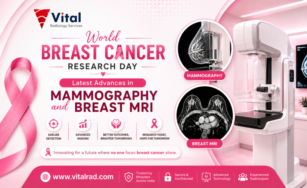

World Breast Cancer Research Day is observed every year on August 18 to raise awareness about ongoing efforts to fight breast cancer. It highlights the importance of research, early detection, and better treatment options for women across the world. Breast cancer remains one of the most common cancers, but early diagnosis can greatly improve survival rates.
Imaging plays a key role in detecting breast cancer at an early stage, often before symptoms appear. Advances in mammography and breast MRI are helping doctors find even small changes in breast tissue more accurately than before.
This article explores the latest developments in these imaging techniques and explains how expert reporting helps ensure timely and precise diagnosis, leading to better patient outcomes.
Why Breast Cancer Research Day Matters
Breast Cancer Research Day is significant, as it puts the spotlight on a disease affecting millions. Recent global statistics reveal breast cancer to be the most common form of cancer globally, with around 2.3 million new breast cancer cases reported annually. In India, breast cancer has become the most prevalent cancer among women, especially in urban centres. Cases of the disease have been rising consistently in recent years, mostly due to lifestyle factors and delays in diagnosis.
Breast Cancer Research Day is not just an event about numbers, but it is dedicated to research, improving people’s access to treatment, and raising awareness about early detection. Many women still delay their health check-ups because of the lack of adequate information, fear, and limited funds. By emphasising the significance of research, Breast Cancer Research Day encourages different stakeholders like governments, the healthcare sector, and communities to invest more in developing better methods for detecting breast cancer.
Research affects the diagnosis and treatment of breast cancer. Over time, technology has improved imaging techniques, such as mammography and breast MRI of the breasts. These improvements have enhanced the accuracy of disease detection, decreased false positives, and contributed to the making of better treatment choices. With the progress of new research, it can eventually provide a chance for earlier diagnosis, less intrusive treatment, and improved women’s survival rates all over the world.
Latest Advances in Mammography
Mammography has improved a lot in recent years. New technologies are helping doctors find breast cancer earlier, even in difficult cases. These advances also make the test faster, safer, and more comfortable for patients.
3D Mammography / Digital Breast Tomosynthesis (DBT)
- A 3D mammogram captures several X-ray pictures of the breast from various positions.
- This allows the physician to identify tiny cancer spots sometimes not visible in 2D assessments.
- This technique is of great use in cases of patients with dense skin tissues, where locating malignancies can be very problematic.
- The use of 3D mammograms boosts success rates of detection of malignancies by 20-40% when compared to the regular techniques.
- The method helps in reducing the number of unnecessary examinations made by women.
- 3D imaging yields better images, thus promoting reliable decisions made by doctors.
AI-Assisted Mammogram Reading
- AI programmes assist radiologists in interpreting mammograms with improved speed and precision.
- These programmes identify potentially abnormal regions, helping radiologists not to overlook significant findings.
- It works like a second pair of eyes, which is important in crowded hospital environments.
- It decreases the chances of false negatives and helps prevent missed cases of cancer.
- Also, it is likely to minimise false-positive cases, reducing patient stress and unnecessary tests.
- Research indicates that in some instances, AI can achieve the accuracy of double readings (The Lancet Digital Health, Nature Medicine).
Contrast-Enhanced Mammography (CEM)
- CEM utilizes a contrasting agent to identify spots with elevated blood flow in the breast. Typically, the cancer cells manifest with an improved blood supply and consequently show up clearer in the images.
- It allows both structural and functional imaging, like MRI.
- It is faster and cheaper than an MRI in several scenarios.
- It comes in handy when a standard mammogram does not produce satisfying results.
- The studies show a sensitivity of over 90% in identifying some types of cancers (American Journal of Roentgenology).
Low-Dose & Faster Screening Protocols
- Current machines use lower levels of radiation without compromising on the quality of images.
- This makes re-staging safer over a longer period of time.
- Faster scanning means shorter waiting time, which improves the comfort of patients.
- Better design means that the discomfort from breast compression becomes less.
- High-speed machines allow screening of more patients in a shorter time.
- All these changes stimulate women to take regular screenings.
Data Snapshot: What Research Shows
- DBT increases detection percentages by as much as 40% while at the same time minimising recall rates by around 15-20% (Radiology).
- AI-powered technologies boost precision and cut down the interpretation time (The Lancet Digital Health).
- CEM shows sensitivity similar to that of MRI, frequently exceeding 90% (AJR clinical studies).
These advances are making mammography more reliable and patient-friendly, helping doctors detect breast cancer early and improve outcomes.
Latest Advances in Breast MRI
Breast MRI is a powerful imaging tool, especially for detecting cancers that may not be visible on mammography. Recent advances have made it faster, more accessible, and more precise, helping doctors diagnose and manage breast cancer with greater confidence.
Abbreviated Breast MRI Protocols
- Abbreviated MRI uses a shorter scanning process compared to traditional MRI.
- It reduces scan time from 30–45 minutes to about 10–15 minutes.
- This makes the test more comfortable and less stressful for patients.
- Lower scan time also reduces cost, making MRI more affordable.
- It allows more patients to be screened in a day, improving access in busy centres.
- Studies show that abbreviated MRI maintains high accuracy, similar to full MRI protocols.
- It is becoming a useful option for screening women with dense breasts.
AI Integration in MRI Interpretation
- AI tools are now helping radiologists read MRI scans more efficiently.
- These systems can detect patterns and highlight areas that may look abnormal.
- AI helps in better lesion characterisation, meaning it can suggest whether a finding looks benign or suspicious.
- It reduces human error and improves consistency between different readers.
- It also saves time by quickly analysing large numbers of images.
- AI can support early diagnosis by identifying subtle changes that may be missed by the eye.
- This technology is still evolving but is already improving diagnostic confidence.
High-Risk Screening Applications
- Breast MRI is strongly recommended for women at high risk of breast cancer.
- This includes women with BRCA1 or BRCA2 gene mutations.
- It is also useful for women with a strong family history of breast cancer.
- MRI works well in dense breast tissue where mammograms may be less effective.
- It helps detect cancers at an earlier stage in high-risk groups.
- Doctors now use a personalised screening approach, combining MRI with mammography based on individual risk.
- This targeted screening improves outcomes and ensures timely treatment.
Improved Contrast Agents & Imaging Resolution
- New contrast agents are being developed to improve image clarity and safety.
- These agents help highlight blood flow patterns, which can indicate cancer activity.
- Improved imaging resolution allows doctors to see smaller and more detailed structures.
- High-quality images help in better tumour detection and accurate staging.
- Advanced MRI machines provide clearer and sharper images than older systems.
- These improvements reduce the chances of missing small lesions.
- Better image quality also supports more precise treatment planning.
These advances are making breast MRI more efficient, accurate, and widely available, helping improve early detection and care for women at all risk levels.
Why Accurate, Timely Reporting Matters
Advanced imaging needs expert reading to deliver the right diagnosis.
- Modern tools like mammography and breast MRI can detect very small changes in breast tissue. However, these images need careful interpretation. A skilled radiologist can understand subtle patterns and decide what is normal and what needs attention. Without expert reading, even the best technology may not give the right answers.
- Subspeciality radiologists who focus only on breast imaging bring deeper knowledge and experience. They are better at identifying early signs of cancer, especially in complex cases like dense breasts. Their expertise helps reduce missed diagnoses and improves overall accuracy.
- Expert interpretation also helps avoid unnecessary biopsies. Not every abnormal-looking area is cancer. A trained specialist can confidently identify harmless changes, which reduces patient stress and avoids extra procedures.
- Timely reporting is just as important as accuracy. Faster report delivery means patients get answers quickly. This reduces anxiety and allows doctors to start treatment without delay when needed.
- In many hospitals, there may not always be access to subspeciality radiologists. Teleradiology helps solve this problem by connecting centres with experienced experts remotely. Images can be shared securely and reviewed by specialists from anywhere.
- This approach improves reporting quality, shortens turnaround time, and ensures that patients receive the right care, no matter where they are.
In simple terms, technology helps detect disease, but expert reporting ensures the right decision is made at the right time.
How Teleradiology Supports Modern Breast Imaging
Teleradiology has greatly improved the quality of breast imaging services around the world, particularly in areas that don’t have many skilled radiologists. This technology lets radiologists observe images remotely, as well as share them safely and securely. Therefore, patients living in rural areas or small hospitals will always have a chance to enjoy professional services without waiting long.
One of the biggest benefits of teleradiology is constant accessibility. Breast imaging procedures, including screenings, generate a large amount of data every day. Nevertheless, timely teleradiology services make it possible to provide timely reports no matter what time of the day it is.
Teleradiology improves the quality of reporting and consistency. The companies in the industry adhere to quality standards and hold ISO certifications, both of which show their commitment to accuracy, safety, and robust processes. Regular audits, peer reviews, and structured reporting systems guarantee that the services delivered meet quality requirements.
Teleradiology combines technology and expertise, thus facilitating timely, precise, and easily accessible breast imaging services. It results in higher quality of diagnosis, faster treatment decisions, and better healthcare services in various types of medical institutions.
The Road Ahead: What’s Next in Breast Imaging Research
Breast imaging is becoming more tailored to each individual’s requirements. Innovative models built on AI technology are coming to the market to estimate the risk of breast cancer in a particular woman. Such tools analyse not only the medical record and imaging results but also the lifestyle data. Doctors will thus be able to form screening strategies based on the data obtained. For example, instead of the standard approach applicable to all women, patients will have a more personalised programme, allowing a woman to be screened for breast cancer more or less frequently or not be screened at all if her particular situation has proved to pose no risk of breast cancer.
Another breakthrough in this area consists of the combination of imaging and genomics data. This will make it possible for physicians to better understand the nature of the disease, thus improving its diagnosis.
Conclusion
Innovations in mammography and breast MRI are revolutionising the process of identifying and treating breast cancer. New technological advances make it possible to capture even the slightest alterations in breast tissues; however, to really add value, the results must be interpreted by qualified specialists. When high-tech imaging is coupled with highly skilled radiology specialists and quick reporting, the outcome is accurate diagnoses that are given on time and contribute to better treatment results for patients.
To obtain reliable and prompt mammogram and breast MRI reports, make use of the services provided by Vital Radiology, where you can find experienced subspeciality radiologists and strict quality assurances.
FAQs
A. What’s the difference between 3D mammography and breast MRI?
3D mammography uses X-rays to create layered images of the breast and is mainly used for routine screening. Breast MRI uses magnetic fields and contrast dye, offering more detailed images, especially useful for high-risk patients or complex cases.
B. Who should get a breast MRI in addition to a mammogram?
Women at high risk, such as those with BRCA1 or BRCA2 gene mutations, a strong family history, prior chest radiation, or very dense breasts, should consider a breast MRI along with a mammogram for more accurate and early detection.
C. How does AI help radiologists in breast imaging?
AI helps radiologists by quickly analysing breast images and highlighting suspicious areas that may need attention. It reduces the chance of missed findings, improves accuracy, and supports faster reporting, especially in busy screening settings with high patient volumes.
D. How often should women get screened?
Screening depends on age and risk level. Most women are advised to start mammograms around 40 and repeat every 1–2 years. High-risk women may need earlier and more frequent screening, sometimes with MRI as recommended.