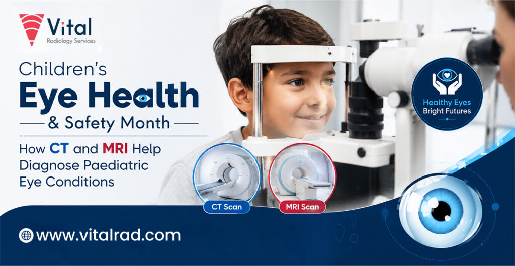

Children’s Eye Health & Safety Month is a time to raise awareness about the importance of protecting children’s vision. Good eye health supports learning, play, communication, and daily activities. It also encourages parents to pay attention to changes in their child’s vision and eye health.
Early diagnosis of eye problems can make a meaningful difference in a child’s development. Some vision problems may not be easy to notice at home, especially when children are young. Regular eye check-ups can help identify concerns early and allow timely treatment. Simple steps, such as protecting the eyes from injury, limiting screen strain, and encouraging healthy habits, can also support better eye health as children grow.
Common Eye Conditions in Children
Most childhood eye problems need only an eye examination, but some conditions require imaging to assess deeper structures and guide care.
- Eye Injuries: Falls, sports accidents, or blunt trauma can damage the eye or surrounding bones. Imaging may help identify fractures, bleeding, or a foreign object.
- Orbital Infections: Infections around the eye can cause redness, pain, fever, swelling, or difficulty moving the eye. Imaging can show whether infection has spread into deeper orbital tissues.
- Congenital Abnormalities: Some children are born with structural problems involving the eye, eye socket, or nearby tissues. Imaging can provide detailed information about these structures and help doctors assess the abnormality.
- Optic Nerve Problems: The optic nerve connects the eye to the brain and carries visual signals. MRI may be recommended when there are unexplained vision changes or concerns about the optic nerve.
- Swelling: Persistent swelling around the eye may be linked to infection, inflammation, injury, or another condition. Imaging can help identify the cause and extent of the swelling.
- Tumours: Tumours involving the eye or surrounding tissues are uncommon in children. Imaging can help doctors determine the location, size, and spread of an abnormal growth and support further treatment planning.
When Is CT or MRI Recommended?
A doctor may recommend CT or MRI when a child has eye symptoms that cannot be fully explained through a routine eye examination. Imaging may be considered after a significant eye injury, especially when there is severe pain, swelling, reduced vision, bleeding, or concern about a fracture or foreign object.
CT may be preferred when doctors need to quickly assess injuries, fractures, or certain infections around the eye. It can provide clear images of the bones and is often useful in urgent situations. MRI may be advised when detailed images of soft tissues are needed, such as the optic nerve, eye muscles, brain, or deeper orbital structures.
Imaging may also be recommended when a child has persistent or unexplained swelling, unusual eye movements, bulging of the eye, sudden vision changes, or suspected congenital abnormalities. MRI can be useful when doctors need to investigate a possible tumour or optic nerve problem. The choice between CT and MRI depends on the child’s symptoms, age, clinical findings, and the suspected condition.
Role of CT in Paediatric Eye Conditions
CT provides detailed images of the eye socket and surrounding bones, helping doctors quickly assess injuries, infections, foreign bodies, and structural changes.
- Identifying Fractures: A strong blow to the face can cause fractures of the thin bones around the eye. CT can show the location and extent of these fractures and help doctors assess nearby structures.
- Assessing Eye Injuries: After a significant injury, CT can help detect damage that may not be visible during an initial examination. It can show changes involving the eye socket, facial bones, and surrounding tissues.
- Detecting Foreign Bodies: Children may accidentally get small objects into or around the eye. CT can help locate certain foreign bodies and determine their position, which can support safe treatment planning.
- Evaluating Infections: Infections around the eye can sometimes spread into deeper orbital tissues. CT can help doctors assess the extent of infection and identify complications, particularly when symptoms are severe or worsening.
- Examining Bone Abnormalities: Some children have congenital or developmental abnormalities involving the bones around the eye. CT provides detailed images of these structures and can help doctors understand their shape and arrangement.
- Supporting Emergency Care: CT is often useful when rapid imaging is needed, particularly after trauma. The scan can provide detailed information quickly, helping doctors make timely decisions about further care.
- Helping Treatment Planning: CT findings can help doctors understand the extent of an injury, infection, foreign body, or bone abnormality. This information may assist specialists in deciding whether observation, medical treatment, or a procedure is appropriate.
Role of MRI in Paediatric Eye Conditions
MRI provides detailed images of soft tissues, helping doctors assess the eye, optic nerve, brain, and visual pathways without radiation.
- Evaluating the Optic Nerve: MRI can provide clear images of the optic nerve and surrounding structures. It may help doctors investigate unexplained vision loss, changes in the optic nerve, or suspected inflammation.
- Examining Eye Muscles: MRI can show the muscles that control eye movement and the tissues around them. It may help evaluate unusual eye movements, restricted movement, or swelling affecting these structures.
- Assessing Soft Tissues: MRI gives detailed images of soft tissues within and around the eye socket. This can help doctors identify inflammation, swelling, structural abnormalities, and other conditions that may not be clearly seen on routine examination.
- Identifying Tumours: When a tumour or abnormal growth is suspected, MRI can help determine its size, location, and relationship with nearby tissues. This information can support diagnosis and treatment planning.
- Checking the Brain and Visual Pathway: Vision involves a pathway connecting the eyes to the brain. MRI can examine the optic nerves, optic chiasm, and relevant brain structures when neurological or visual symptoms raise concern.
- Assessing Congenital Conditions: MRI can help doctors evaluate certain conditions present from birth by providing detailed views of the eye, orbit, optic nerves, and brain structures. The findings can support further specialist assessment and care.
CT vs MRI: Which One Is Used?
CT may be preferred when a child needs quick imaging after an eye or facial injury. It provides detailed images of the bones around the eye and can help identify fractures, certain foreign bodies, bleeding, and changes linked to some infections. Because CT is fast, it can be useful in urgent situations. However, it uses ionising radiation, so doctors consider the child’s age and the expected benefit before recommending it.
MRI may provide better information when doctors need detailed images of soft tissues. It can show the optic nerve, eye muscles, orbital tissues, brain, and visual pathways. MRI may be useful when investigating optic nerve problems, tumours, congenital abnormalities, or unexplained changes affecting vision. It does not use ionising radiation, but the scan takes longer, and children may need help staying still.
The final choice depends on the child’s symptoms and clinical findings.
Safety of CT and MRI in Children
CT uses ionising radiation to create detailed images, so doctors carefully consider its need in children. Since children are more sensitive to radiation, unnecessary CT scans should be avoided. When a CT is needed, the imaging team uses an appropriate, child-specific dose to obtain useful images while limiting radiation exposure.
MRI does not use ionising radiation and uses magnetic fields and radio waves to produce detailed images. It can be useful for examining soft tissues, the optic nerve, and visual pathways. However, MRI may take longer and require the child to remain still. Both tests should be performed only when clinically appropriate and recommended by a qualified doctor.
Importance of Accurate Radiology Reporting
Accurate radiology reporting helps doctors understand what CT or MRI shows and how the findings may relate to a child’s symptoms. A clear report can identify important abnormalities, describe their location and extent, and provide useful information for further evaluation.
Timely reporting is especially important when a child has an injury, infection, sudden vision changes, or another urgent concern. Reliable imaging interpretation helps the treating doctor make informed decisions about further tests, treatment, monitoring, or specialist care. It also supports clear communication among healthcare professionals.
Conclusion
Protecting a child’s vision starts with noticing changes early and seeking medical advice when something seems unusual. Symptoms such as persistent swelling, eye injuries, vision changes, or unusual eye movements should not be ignored. Early evaluation can help identify problems before they affect a child’s daily life or development.
CT and MRI should be used only when clinically appropriate and when their findings can add useful information. Accurate imaging and timely reporting help doctors choose suitable care. Parents can support healthy vision by scheduling recommended eye checks and following medical advice.