
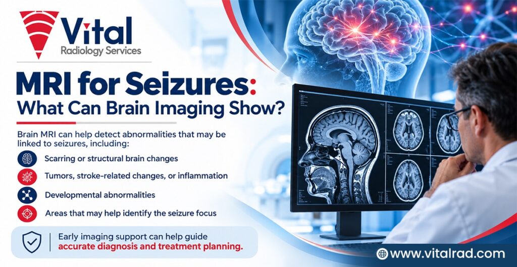

A first seizure can be concerning, particularly when there is no clear explanation for what caused it. If seizures happen repeatedly or remain unexplained, doctors may recommend a brain MRI as part of the evaluation. MRI creates detailed images of the brain and can help identify structural changes that may be linked to seizure activity.
During the scan, radiologists carefully assess the brain for abnormalities such as developmental differences, scars, tumours, blood-vessel changes, or damage from an earlier injury. MRI may also provide useful information about the location and appearance of these changes.
This article looks at why MRI is used in seizure evaluation, what brain imaging can reveal, and how the findings may support further assessment and care.
Why Is MRI Important in the Evaluation of Seizures?
A brain MRI can provide important information about possible structural causes of seizures:
- Identifies brain abnormalities: MRI can detect structural changes, such as scars, tumours, developmental abnormalities, or areas of previous brain injury that may be linked to seizures.
- Detects small lesions: Detailed MRI images can reveal some abnormalities that may not be clearly visible on other imaging tests.
- Helps locate seizure-related changes: MRI findings can help neurologists understand where a structural abnormality is located and whether it could be related to seizure activity.
- Important for focal seizures: MRI is particularly useful when seizures appear to begin in one specific area of the brain.
- Useful in recurrent seizures: When seizures continue or their cause remains unclear, MRI may help identify an underlying structural reason.
- Supports epilepsy assessment: MRI is especially important when epilepsy is difficult to control despite medicines.
However, MRI is only one part of the evaluation. Doctors also consider the person’s medical history, symptoms, neurological examination, and EEG findings.
MRI vs CT Scan for Seizures: What Is the Difference?
MRI and CT scans are both useful brain imaging tests, but they serve different purposes when investigating seizures.
|
Feature |
MRI |
CT Scan |
|
Imaging detail |
Provides detailed images of brain tissue and structure |
Provides clear images quickly, particularly for major abnormalities |
|
Radiation exposure |
Does not use ionising radiation |
Uses ionising radiation |
|
Scan time |
Usually takes longer |
Usually takes only a few minutes |
|
Emergency use |
Less commonly used for immediate emergency imaging |
Often preferred when rapid imaging is needed |
|
Subtle brain abnormalities |
Better at detecting many small or subtle structural changes |
May not detect some subtle abnormalities |
| Common clinical applications | Epilepsy evaluation and detailed assessment of brain structure |
Emergency assessment, head injury and suspected bleeding |
CT is often used when doctors need quick brain imaging, particularly after a head injury or when bleeding, swelling, or another urgent problem is suspected. It may also be used when MRI is not immediately available or cannot be performed. MRI is generally preferred for detailed evaluation of recurrent or focal seizures. It can provide a closer look at subtle structural changes that may help doctors understand the possible cause of seizures.
What Is an Epilepsy-Protocol MRI?
An epilepsy-protocol MRI is a specialised brain scan designed to identify structural changes associated with seizures. Unlike a routine MRI, it uses specific sequences and carefully planned views of brain regions commonly involved in epilepsy. Thin-slice imaging, 3D T1 and FLAIR sequences, and coronal T2 images focused on the hippocampus can provide detailed views of brain tissue and help detect subtle abnormalities.
A 3T MRI can offer high-resolution images and may improve the detection of small lesions. Routine MRI may miss subtle epileptogenic abnormalities because of differences in image resolution, sequences, or orientation. An epilepsy-specific protocol provides a more focused assessment when epilepsy is suspected.
What Can a Brain MRI Show in Patients With Seizures?
A brain MRI can help identify several structural abnormalities that may be linked to seizures and provide clues about their possible cause.
Mesial Temporal Sclerosis
Mesial temporal sclerosis involves shrinkage (volume loss) and abnormal MRI signal in the hippocampus. It is commonly associated with temporal lobe epilepsy and can be particularly important when evaluating drug-resistant epilepsy.
Focal Cortical Dysplasia
Focal cortical dysplasia develops when the brain’s outer layer (cortex) does not develop normally. These changes can be very subtle and may be difficult to detect on routine MRI. Dedicated epilepsy protocols and review by an experienced neuroradiologist can improve detection.
Brain Tumours
Some brain tumours, particularly slow-growing or low-grade tumours, may first present with seizures. MRI can help show the tumour’s location, size and imaging characteristics, helping doctors understand its relationship with nearby brain structures.
Neurocysticercosis and Brain Infections
Neurocysticercosis is an important cause of seizures in areas where the infection is common, including India. MRI may show cysts, surrounding swelling (oedema), enhancement, or other changes associated with infection.
Vascular Abnormalities
Cavernous malformations, arteriovenous malformations and other blood-vessel abnormalities can sometimes be associated with seizures. MRI can help identify these structural changes and assess their location.
Stroke, Trauma and Gliosis
A previous stroke, head injury, or other brain injury can leave areas of permanent structural damage or gliosis (scar-like brain tissue). These areas may later become associated with seizures.
Developmental and Genetic Conditions
MRI can also help identify developmental abnormalities linked to epilepsy, including conditions such as tuberous sclerosis. Detailed imaging can be especially useful when evaluating children with epilepsy.
What Does a Normal MRI Mean in Seizure Evaluation?
A normal MRI does not always mean that seizures or epilepsy are absent. The scan is only one part of the overall assessment.
- A normal MRI does not rule out epilepsy: Many people with epilepsy may have no visible structural abnormalities on brain imaging.
- Some epilepsies do not cause detectable changes: Certain seizure disorders occur without obvious changes in brain structure.
- Some lesions can be very subtle: Small abnormalities may be difficult to identify, even on detailed imaging.
- Clinical information remains important: MRI findings are interpreted along with medical history, seizure symptoms (semiology), neurological examination and EEG results.
- Advanced imaging may sometimes be considered: In selected cases, doctors may recommend repeat MRI, epilepsy-protocol MRI or other specialised imaging tests.
- Expert review can make a difference: Specialist neuroradiologists with experience in epilepsy imaging may identify subtle findings that could be missed on routine review.
- Seizure evaluation is a combined process: Doctors use information from multiple tests to better understand the possible cause of seizures and plan further care.
Why Does Specialist Neuroradiology Reporting Matter in Epilepsy MRI?
Careful interpretation of an epilepsy MRI can help identify subtle findings that may otherwise be overlooked.
- Looks for subtle abnormalities: Epilepsy MRI requires careful attention to small structural changes that may be associated with seizure activity.
- Helps detect difficult-to-see lesions: Findings such as focal cortical dysplasia or mild hippocampal abnormalities can be subtle and may not be obvious on routine review.
- Uses a systematic approach: Experienced neuroradiologists assess specific brain regions and MRI sequences systematically, particularly areas commonly involved in epilepsy.
- Allows comparison with earlier scans: When previous imaging is available, comparing examinations can help identify subtle changes or clarify findings that were previously uncertain.
- Provides clinically useful information: A detailed radiology report describes relevant abnormalities, their location and other imaging features, giving the treating neurologist information that can support clinical decision-making.
- Supports complex epilepsy evaluation: Specialist interpretation can be particularly valuable when seizures continue despite treatment or when the cause remains unclear.
- Important for surgical planning: In selected patients being assessed for epilepsy surgery, accurate identification and localisation of structural abnormalities can contribute to the multidisciplinary evaluation and surgical planning process.
How Vital Radiology Supports Seizure and Epilepsy MRI Reporting
Accurate MRI interpretation can provide valuable information during seizure evaluation, particularly when brain abnormalities are subtle or difficult to identify.
Specialist Image Interpretation
Vital Radiology supports complex brain MRI reporting through specialist image interpretation. Neuroradiologists review the images carefully, considering the clinical information and MRI sequences to identify structural abnormalities that may be associated with seizures or epilepsy.
Attention to Subtle Findings
Some seizure-related abnormalities can be difficult to detect on routine review. Specialist assessment can focus on findings such as focal cortical dysplasia, hippocampal sclerosis, and small vascular lesions. Careful review may help ensure that subtle imaging features are appropriately recognised and documented.
Structured Clinical Reporting
A clear and structured report can make imaging findings easier for the treating team to understand. Relevant abnormalities, their location, and important imaging characteristics can be described clearly, providing the referring neurologist or other clinician with useful information for further evaluation and treatment planning.
Teleradiology Support
Teleradiology allows medical images to be reviewed remotely by qualified radiologists. Imaging studies can be securely shared for interpretation, with reports delivered digitally to healthcare providers. This approach can support access to specialist radiology expertise across different healthcare settings.
Epilepsy vs Seizures: What Is the Difference?
A seizure is an episode of abnormal electrical activity in the brain, while epilepsy is a neurological condition involving a tendency to have recurrent unprovoked seizures.
|
Features |
Seizure |
Epilepsy |
|
Definition |
A single episode of abnormal brain activity |
A condition associated with recurrent unprovoked seizures |
|
Causes |
May result from fever, injury, infection or other triggers |
May have genetic, structural or unknown causes |
|
Frequency |
Can occur once |
Involves an ongoing risk of seizures |
|
Diagnosis |
Based on symptoms and clinical assessment | May involve history, EEG, MRI and other tests |
|
MRI |
May identify an underlying cause |
Helps look for structural abnormalities |
|
EEG |
May detect abnormal electrical activity |
Helps support epilepsy diagnosis and classification |
Doctors evaluate suspected epilepsy by reviewing the seizure history, symptoms, and neurological examination. EEG may assess brain electrical activity, while MRI can look for structural changes that could be linked to seizures.
When Can Additional Imaging Review Be Valuable?
Additional imaging review can be valuable when MRI findings are subtle, difficult to characterise, or reported as indeterminate or non-specific. It may also be considered when there is a mismatch between the clinical or EEG findings and the initial MRI interpretation. Reviewing previous scans can provide useful comparison and help clarify uncertain findings.
Further review may be particularly useful when seizures continue despite treatment or when a person is undergoing a complex epilepsy evaluation. A detailed assessment can also be important during epilepsy surgery evaluation, where understanding the location and characteristics of possible seizure-related abnormalities may contribute to clinical decision-making.
Specialist Neuroradiology Reporting With Vital Radiology
Specialist interpretation can add valuable expertise when brain MRI findings are subtle, complex, or require careful clinical correlation.
- Specialist teleradiology support: Vital Radiology provides remote radiology reporting support, helping healthcare providers access specialist imaging expertise without requiring the radiologist to be physically present at the imaging facility.
- Experienced neuroradiologists: Complex brain MRI examinations can be reviewed by experienced neuroradiologists who understand the imaging features relevant to seizures and epilepsy.
- Attention to subtle findings: Careful image review focuses on structural abnormalities that may be difficult to recognise, including focal cortical dysplasia, hippocampal abnormalities and small vascular lesions.
- Clinical correlation: Where available, relevant clinical history and previous imaging can be considered during image interpretation. This information can help place MRI findings in the appropriate clinical context and support comparison with earlier examinations.
- Clear, clinically relevant reporting: Reports can describe important findings, their location and relevant imaging characteristics in clear language, giving referring doctors and healthcare providers information that can support further evaluation and management.
- Support for complex evaluations: Specialist neuroradiology reporting may be useful when MRI findings are uncertain, seizures persist despite treatment, or imaging forms part of a broader epilepsy assessment.
- Focus on dependable reporting: Vital Radiology focuses on specialist neuroradiology reporting designed to provide consistent, clinically relevant interpretation and support healthcare teams in their decision-making.
Conclusion
Brain MRI can provide valuable information when evaluating seizures by identifying structural abnormalities that may be linked to seizure activity. An epilepsy-protocol MRI and careful neuroradiology review can be particularly useful when findings are subtle or when seizures continue despite treatment. However, MRI is only one part of the evaluation and should be considered alongside clinical history, seizure characteristics and EEG findings.
For specialist interpretation of seizure and epilepsy-related MRI studies, connect with Vital Radiology for expert teleradiology reporting support. Specialist review can help provide clear, clinically relevant imaging information to support neurologists and healthcare providers during further evaluation and management.
Frequently Asked Questions
- Can MRI detect the cause of seizures?
MRI can identify structural abnormalities associated with seizures, including scars, tumours, developmental changes and vascular lesions. However, some people with epilepsy have no visible structural abnormality, so MRI may not identify the cause in every patient.
- Is MRI or CT better for evaluating seizures?
MRI and CT have different roles in seizure evaluation. CT is commonly used for rapid emergency assessment, while MRI provides greater soft-tissue detail and is generally better for identifying many structural abnormalities associated with epilepsy.
- How long does an epilepsy MRI take?
The duration of an epilepsy MRI can vary depending on the imaging protocol, number of sequences, and imaging centre. A dedicated epilepsy protocol may take longer than a routine brain MRI because it requires additional, detailed images.
- Can an MRI miss the cause of seizures?
Yes, an MRI can sometimes miss the cause of seizures. Small lesions may be hard to see. Some epilepsy has no visible changes. A routine MRI may also miss findings detected with a dedicated epilepsy protocol.
- Is MRI safe for children with seizures?
MRI is generally safe for children with seizures and does not use ionising radiation. However, young children may need extra preparation or sedation if they cannot stay still during the scan. The treating team will assess their needs beforehand.
- Why is neuroradiology expertise important for epilepsy MRI?
Neuroradiology expertise can help identify subtle brain abnormalities that may be linked to seizures. A specialist also considers the clinical question and relevant history when reviewing MRI findings, helping provide a clearer and more useful interpretation.
- Can a radiology report provide a diagnosis of epilepsy?
A radiology report does not diagnose epilepsy on its own. The radiologist interprets brain imaging findings, while the treating team considers medical history, examination, EEG, and other relevant tests to make the diagnosis and guide further care.