A sudden headache, blurred vision, or unexplained dizziness; are these harmless symptoms or warning signs of something far more serious? The line between a migraine and a life-threatening neurological condition can be dangerously thin. What seems minor today could silently progress into irreversible damage if overlooked.
Early detection plays a critical role in preventing severe complications such as stroke-related disability or tumor growth. Delayed diagnosis increases the risk of permanent brain injury, affecting quality of life and recovery outcomes. Timely neuroradiology reporting not only clarifies the cause but also accelerates crucial treatment decisions, ensuring patients receive the right care when it matters most.
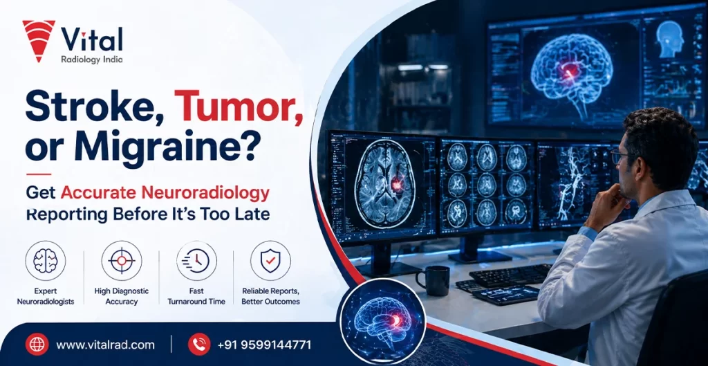
Understanding the Difference: Stroke vs Tumor vs Migraine
Understanding neurological symptoms can be challenging, as conditions like stroke, brain tumors, and migraines often share overlapping signs. The table below highlights key differences to help clarify their nature, symptoms, and urgency.
| Condition | Definition | Common Symptoms | Onset | Severity Level | Imaging |
| Stroke | Sudden interruption of blood flow to the brain, either due to blockage or bleeding | Sudden weakness (especially one side), slurred speech, confusion, vision problems, loss of balance | Very sudden, often without warning | Medical emergency requiring immediate attention | Urgent imaging (CT/MRI) is critical to confirm type and begin life-saving treatment |
| Brain Tumor | Abnormal and uncontrolled growth of cells within the brain | Persistent headaches, seizures, personality changes, vision or speech difficulties, nausea | Gradual onset, symptoms worsen over time | Potentially life-threatening depending on type and size | Detailed imaging helps detect the location, size, and nature of the tumor |
| Migraine | A neurological condition causing intense, recurring headaches, often with sensory disturbances | Throbbing headache, nausea, sensitivity to light/sound, visual aura, dizziness | Develops gradually, may have triggers | Usually not life-threatening, but can be debilitating | Imaging may be needed to rule out other serious conditions if symptoms are unusual |
Accurate brain imaging diagnosis is essential to distinguish between these conditions, ensuring timely and appropriate treatment while preventing misdiagnosis and complications.
Common Symptoms That Should Never Be Ignored
Certain neurological symptoms may appear mild at first but can signal serious underlying conditions that require urgent medical attention. Recognizing these warning signs early and seeking timely neuroimaging can be life-saving.
- Persistent severe headache or worsening pain should never be dismissed as a routine issue. An unusually intense headache that lasts longer than expected or progressively worsens over time may indicate conditions such as internal bleeding, increased intracranial pressure, or a growing mass in the brain.
- Sudden vision problems, dizziness, or numbness are critical warning signs that demand immediate evaluation. Blurred or double vision, a spinning sensation, or numbness in the face, arms, or legs, especially on one side, can point toward disrupted brain function, often associated with stroke or other neurological disorders.
- Difficulty in speech or loss of coordination is a serious red flag that requires urgent care. Slurred speech, inability to find words, confusion, or trouble maintaining balance and coordination may indicate impaired brain activity, which could rapidly worsen without prompt diagnosis and treatment.
These symptoms require immediate neuroimaging tests to identify the root cause accurately. Techniques such as CT scans or MRI play a crucial role in detecting abnormalities early, enabling faster medical intervention and significantly improving patient outcomes.
Role of Neuroradiology in Accurate Diagnosis
Neuroradiology plays a crucial role in modern medicine by helping detect abnormalities within the brain that may not be visible through routine clinical examinations. Advanced imaging techniques such as MRI and CT scans allow specialists to identify issues like bleeding, blockages, or abnormal tissue growth at an early stage, enabling timely intervention.
It also provides clear and detailed insights into both the structure and function of the brain. High-resolution imaging helps doctors assess the exact location, size, and extent of any abnormality while also evaluating how different regions of the brain are functioning, leading to more accurate clinical decisions.
Most importantly, neuroradiology reporting helps doctors differentiate between conditions such as stroke, brain tumors, and migraines, which often present with similar symptoms. This clarity ensures patients receive the right treatment promptly, reducing the risk of complications and improving overall outcomes.
Advanced Imaging Techniques Used in Neuroradiology
Modern neuroradiology relies on advanced imaging technologies to provide precise and timely insights into brain health. These techniques are essential for accurate diagnosis, early detection, and effective treatment planning.
- MRI Scan (Magnetic Resonance Imaging): Is widely used for capturing detailed images of soft tissues in the brain. It helps identify tumors, inflammation, nerve damage, and other subtle abnormalities that may not be visible with other imaging methods.
- CT Scan (Computed Tomography): Is highly effective for rapid assessment in emergencies. It is commonly used to quickly detect internal bleeding, skull fractures, or acute stroke, making it crucial for immediate medical decision-making.
- Functional MRI (fMRI): Is used to analyze brain activity by measuring changes in blood flow. It helps map functional areas of the brain, supporting diagnosis and planning for surgeries or neurological treatments.
Diffusion imaging plays a key role in detecting early signs of stroke. It identifies restricted blood flow at a very early stage, allowing prompt intervention and reducing the risk of permanent brain damage.
How Stroke is Diagnosed Through Imaging
Timely imaging plays a life-saving role in diagnosing stroke, as it helps determine the type, severity, and exact location of the problem, enabling immediate and appropriate treatment decisions.
CT Scan for Instant Bleeding Detection
- A CT scan is typically the first imaging test used in suspected stroke cases due to its speed and wide availability.
- It quickly detects bleeding in the brain, making it essential for diagnosing hemorrhagic stroke. Early identification ensures that patients receive the correct emergency treatment without delay, which can significantly reduce the risk of severe complications or death.
MRI Brain Scan for Early Ischemic Stroke
- An MRI brain scan is highly sensitive in identifying ischemic stroke, which is caused by a blockage in blood flow.
- It can detect subtle changes in brain tissue at a very early stage, often before they become visible on a CT scan. This allows doctors to confirm the diagnosis and initiate targeted therapies to restore blood flow and minimize brain damage.
Rapid neuroradiology reporting is critical in emergency stroke care.
Quick and accurate interpretation of imaging results helps doctors make immediate treatment decisions, such as administering clot-busting drugs or planning surgical intervention, ultimately improving patient outcomes and survival rates.
Detecting Brain Tumors with Precision
Detecting brain tumors with precision relies heavily on advanced imaging techniques that provide detailed information about the tumor’s size, location, and characteristics. These insights help clinicians understand the nature of the growth and assess its potential impact on surrounding brain structures.
Accurate imaging also plays a critical role in planning treatment, including surgery or radiation therapy. By clearly defining the tumor, doctors can choose the most effective approach while minimizing risks. Early and precise diagnosis significantly improves treatment outcomes and enhances the chances of successful recovery.
Identifying Migraine Through Neuroimaging
Neuroimaging plays an important role in identifying migraines, especially when symptoms overlap with more serious neurological conditions. While migraines themselves may not always show visible changes on scans, imaging helps rule out critical issues such as brain tumors or stroke that can present with similar warning signs.
It also supports diagnosis when symptoms are unclear, severe, or unusual in pattern. By eliminating other possible causes, doctors can confidently confirm migraines and design an appropriate treatment plan. This ensures patients receive targeted care, avoid unnecessary interventions, and manage their condition more effectively over time.
Importance of Timely Neuroradiology Reporting
Timely neuroradiology reporting is critical in ensuring that patients receive accurate diagnoses and immediate medical attention, especially in urgent neurological conditions where every second matters.
- Faster reports lead to quicker medical intervention and improved patient outcomes. When imaging results are delivered promptly, doctors can make swift decisions regarding treatment, reducing delays that could worsen the patient’s condition.
- Early reporting significantly reduces the risk of complications and long-term disability. Conditions affecting the brain can progress rapidly, and any delay in diagnosis may result in irreversible damage. Timely reports help initiate appropriate therapies at the earliest possible stage.
- It is especially essential in emergency cases such as acute stroke. Rapid neuroradiology interpretation allows clinicians to determine the type of stroke and begin life-saving treatments, such as clot removal or bleeding control, within critical time windows.
Efficient reporting enhances overall care coordination. It ensures seamless communication between radiologists and treating physicians, leading to accurate diagnosis, better treatment planning, and improved recovery chances.
Risks of Misdiagnosis Without Proper Imaging
Without proper neuroimaging, serious neurological conditions can be easily misdiagnosed, leading to delayed or inappropriate treatment and increased health risks.
- Stroke may be mistaken for a migraine, delaying critical treatment. This confusion can prevent timely intervention, increasing the risk of permanent brain damage or disability.
- Brain tumors may go undetected without advanced imaging techniques. Subtle or early-stage growths often require detailed scans for accurate identification.
- Misdiagnosis significantly increases the risk of severe health complications. Delayed or incorrect treatment can worsen the condition, reduce recovery chances, and impact overall quality of life.
Choosing the Right Diagnostic Center for Neuroradiology
Choosing the right diagnostic center for neuroradiology is essential for accurate diagnosis and timely treatment. It is important to select a facility equipped with advanced imaging technologies such as MRI and CT scans, ensuring precise detection of neurological conditions.
Additionally, the presence of experienced radiologists and efficient reporting systems plays a crucial role. Fast and accurate reports help doctors make prompt decisions, especially in emergency cases, ultimately improving patient outcomes and reducing the risk of complications.
When Should You Seek Neuroradiology Testing?
Knowing when to seek neuroradiology testing is essential for early diagnosis and preventing serious neurological complications. Paying attention to warning signs can help ensure timely medical evaluation and treatment.
- Sudden onset of neurological symptoms should never be ignored. Symptoms such as weakness, numbness, confusion, vision changes, or difficulty speaking may indicate a serious condition like stroke and require immediate imaging.
- Chronic headaches that do not improve over time need proper evaluation. Persistent or worsening headaches, especially those that differ from usual patterns, may signal underlying neurological issues that require detailed imaging.
- A history of brain disorders or head injuries increases the need for monitoring. Individuals with previous neurological conditions or trauma may require periodic imaging to detect any changes or complications early.
- Doctor-recommended brain imaging tests should always be taken seriously. If a healthcare provider advises neuroimaging, it is crucial to follow through promptly to ensure accurate diagnosis and appropriate treatment planning.
Conclusion
Accurate neuroradiology reporting helps doctors clearly understand what is happening in the brain. Many serious conditions can look similar at first, but proper imaging makes the diagnosis correct. Taking action early can prevent major problems like brain damage and long-term health issues.
Do not wait or ignore symptoms that feel unusual. If your doctor suggests a scan, get it done without delay. Quick testing and fast reports help start the right treatment sooner. Making timely decisions can protect your health and improve recovery in a simple but powerful way.