
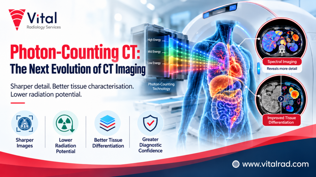

CT scans allow medical professionals to observe the body in order to detect diseases and conditions. As technology progresses in the world of imaging, photon-counting CT offers many benefits in health care. Unlike traditional CT scanners, which use conventional methods, this new technology captures individual X-ray photons along with their energy to produce high-definition images of the interior of the body.
Such detailed imaging can produce more accurate results when it comes to spotting minor anomalies and evaluating medical conditions. Furthermore, this technology is more likely to decrease the amount of radiation that the patient is exposed to. From detection of cardiovascular diseases to cancers, photon-counting CT will help to make better diagnoses and provide a high-quality level of care.
What Makes Photon-Counting CT Different
Photon-counting CT is an advanced imaging technology that differs from conventional CT scanners in how it detects and processes X-rays. The key differences include:
- Direct photon detection: Unlike conventional CT scanners, which first convert X-rays into light, photon-counting CT detectors measure individual X-ray photons directly.
- Better image detail: This technology can produce sharper images, helping doctors examine small structures and identify subtle abnormalities.
- Energy measurement: It measures the energy of individual photons, providing additional information about the tissues being examined.
- Improved tissue differentiation: The scanner can help distinguish between different materials, supporting more detailed assessments of certain conditions.
- Potentially lower radiation exposure: It can reduce radiation doses in some examinations while maintaining useful image quality.
- Reduced image noise: Advanced detection can improve image clarity, particularly when examining small anatomical structures.
- Smaller detector elements: These can capture finer details and improve the visualisation of tiny structures, including blood vessels.
These features may support more accurate diagnoses and better-informed treatment decisions.
What the Technology Improves
Photon-counting computed tomography (CT) provides many advantages over traditional CT imaging. It captures detailed information from individual X-ray photons, allowing physicians to see clearer images of internal structures. Significant improvements include the following:
- Image quality: It provides sharper images that enable detection of tiny lesions and examination of fine anatomical structures.
- Radiation dose: It enables a reduction in radiation exposure during specific studies while retaining good image quality.
- Tissue assessment: By employing energy-based imaging, it allows identification of different tissues and materials, providing additional diagnostic information.
- Reduced image noise: This technique can enhance the quality of images by minimising the unwanted visual noise that obscures details.
- Heart imaging: It enables visualisation of small blood vessels and evaluation of coronary arteries, including in selected patients with significant calcification.
- Cancer evaluation: Excellent quality images allow one to examine tumours and surrounding areas with greater efficiency.
- Confidence in diagnosis: Improved image quality and additional information about tissues allow for better-informed and more confident clinical decisions.
These innovations help radiologists obtain helpful diagnostic data as well as adapt examinations to the specific needs of individual patients.
Where Photon-Counting CT Is Being Used Clinically
Photon-counting CT enhances medical imaging in numerous fields by giving quality images and important diagnostic insights.
Cardiovascular Imaging
With this technology, physicians can examine the heart and vessels in a clearer manner. High-quality images from photon-counting CT are useful for assessing coronary arteries even when obstructed by calcification. As a result, it can be used to judge coronary artery disease and decide how treatment protocols should be designed.
Cancer Detection and Assessment
Photon-counting CT is also able to capture images that enable doctors to analyse tumours and tissues surrounding them. By being able to tell materials apart based on energy characteristics, the technology can yield a lot of relevant information regarding various anomalies.
Lung Imaging
This technology captures precise details in lung structure, which can help examine small nodules and other issues. What is more, it can assist in the assessment of certain lung diseases thanks to high image quality.
Bone and Joint Imaging
Photon-counting CT makes it possible to obtain images of bones, joints, and very small fractures. With its high spatial resolution, this technology can be employed to analyse complicated structures of bone and certain conditions of orthopaedics.
Brain and Head Imaging
This technology can improve the visualisation of fine structures in the head. Depending on the clinical indication, it may support the assessment of selected neurological conditions. As clinical experience grows, photon-counting CT may offer further benefits across diagnostic imaging.
Where the Technology Stands in 2026
By 2026, photon-counting CT has moved beyond research into clinical use, with hospitals adopting the technology for a growing range of diagnostic examinations. Recent advances are expanding its applications across cardiac, lung, brain, abdominal, and musculoskeletal imaging.
New scanner systems are also receiving regulatory clearances, helping expand access to this technology in different markets. These developments reflect growing interest in clearer images, improved tissue assessment, and more efficient scanning.
However, availability remains limited compared with conventional CT, and costs, workflow changes, and the need for further clinical evidence may affect adoption. Researchers continue to study how best to use the technology in routine patient care.
What Photon-Counting CT Means for Reporting
Photon-counting CT provides detailed images, requiring careful interpretation and structured reporting.
- More image details: Higher image resolution allows radiologists to examine smaller structures and subtle abnormalities. Reports may need precise descriptions of findings to support accurate clinical decisions.
- Additional tissue information: Energy-based measurements can help distinguish certain materials. Radiologists may need to include these findings when they provide useful diagnostic information.
- Improved coronary assessment: Detailed images of coronary arteries may help radiologists describe small vessels, calcium deposits, and other features relevant to cardiovascular disease.
- Clearer findings: Reduced image noise can improve visibility in some examinations, helping radiologists assess abnormalities and communicate their significance more clearly.
- Adapted reporting protocols: Departments may need to update reporting guidelines to reflect the scanner’s capabilities and ensure consistency across examinations.
- Ongoing training: Radiologists require appropriate training to interpret spectral information and understand the clinical relevance of photon-counting CT findings.
- Clinical collaboration: Clear communication between radiologists and referring doctors helps ensure that additional imaging information supports patient management.
What Is Still Unresolved
Although photon-counting CT offers promising advances in medical imaging, several challenges need to be addressed before it becomes widely available in routine clinical practice.
- High equipment costs: Photon-counting CT scanners require significant investment, which may limit adoption in smaller hospitals and healthcare facilities.
- Limited availability: Access remains restricted in many regions, meaning patients may not benefit from this technology locally.
- Need for further research: More studies are needed to establish how consistently the technology improves diagnosis and patient outcomes across different conditions.
- Training requirements: Radiologists and technicians need specialised training to understand the technology and interpret its findings accurately.
- Reporting standards: Healthcare facilities need clear reporting guidelines to ensure consistent interpretation and communication of findings.
- Clinical integration: Hospitals must adapt existing workflows and protocols to incorporate the technology effectively.
An Evolution Already Underway
The development of photon-counting computed tomography (CT) is a significant advancement in the field of medical imaging. This technology allows for the detection and measurement of individual X-ray photons, which leads to the generation of high-quality imaging data. These advancements may aid radiologists in their endeavors to identify various health conditions in patients.
Although hospitals are actively searching for practical uses for photon-counting CT, it is being utilized in radiology departments, cardiology, oncology, and pulmonary studies as well. The key factors that will contribute to the successful implementation of photon-counting CT technology are affordability, training, and proof of its advantages.
For radiologists, the goal is to turn improved imaging data into useful clinical data. Further research may make photon-counting CT very useful in modern diagnostic imaging.
How Vital Radiology Helps
Advancements in the field of medical imaging continue to make quality and timely reporting an integral component of efficient patient treatment. Vital Radiology provides reputable radiology reporting services to healthcare professionals that are flexible enough to fit specific clinical applications. Additionally, the support of a reporting partner enables the effective management of both regular and abnormal volumes of requests while ensuring prowess in both reporting efficiency and consistency.
Discover how Vital Radiology can be utilized to accommodate the requirements of imaging teams adapting to the latest processes in diagnostic imaging.
Conclusion
Photon-counting CT technology stands as a significant advancement in the field of medical imaging, delivering sharper images and more comprehensive data about tissues within the body. Its use in diagnosing heart and lung diseases and fighting cancer illustrates its potential for improving the diagnosis and treatment of illnesses. Nonetheless, the future use of this technology will depend on its cost and proper training of personnel, as well as additional medical studies.
As photon-counting CT keeps improving, radiologists will have to find out how this technology may be beneficial for patients. Understanding its advantages and limits will allow hospitals to choose the best approach to using the technology for imaging.