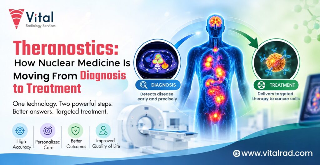

Nuclear medicine has traditionally enabled doctors to detect diseases by means of small doses of radioactive materials to take pictures of organs and tissues. Nowadays, it has branched out from being just a diagnostic tool to theranostics that combine diagnosis and therapy in one.
In theranostics, radioactive drugs are used that can identify specific targets in abnormal cells and deliver treatment whenever possible. The first scan helps to determine if the target is present in the body, and if it is, the treatment drug is administered. Thus, it enables more targeted treatment and better control of the progress of the disease.
What Are Theranostics in Nuclear Medicine?
Theranostics is a medical approach that combines diagnosis and treatment. It uses radioactive medicines that can find specific targets on abnormal cells. A diagnostic scan is first used to see if these targets are present.
If the target is found, a similar radioactive medicine can be used for treatment. It carries radiation directly to the targeted cells. This may help doctors treat disease more precisely while reducing the effect on nearby healthy cells. Theranostics is mainly used for some cancers and is being studied for other conditions.
What Are the Challenges of Theranostics?
Although theranostics offer a targeted approach to diagnosis and treatment, their use can involve several practical challenges.
-
Patient Selection
Not every patient is suitable for treatment. Doctors first need to confirm that the disease has the specific target that the radioactive medicine can recognise. Accurate imaging and other clinical information help determine whether a patient may benefit from treatment.
-
Availability of Radiopharmaceuticals
Theranostics depend on specialised radioactive medicines called radiopharmaceuticals. Their availability may vary between hospitals and regions. Short supply, production needs and transport requirements can sometimes delay access to treatment.
-
Specialised Knowledge and Equipment
Theranostics requires professional nuclear medicine specialists, medical physicists, radiopharmacists, and other medical employees. Hospitals require special equipment for carrying out imaging procedures, treatment, and protection against radiation. Additionally, these requirements might pose obstacles for access to this method in some centres.
-
Importance of Clear Imaging and Reporting
It is necessary to carry out quality imaging and reporting when selecting patients or planning treatment. Doctors must understand where the target is located and how far the disease has spread. Also, consistent reporting makes it possible for the medical team to monitor treatment efficiency.
How Does the Theranostic Approach Work?
The theranostic approach follows a step-by-step process that helps doctors identify suitable patients, deliver targeted treatment and check how well it is working.
-
Identifying Molecular Targets
The process begins by identifying a specific molecular target on abnormal cells. These targets may be proteins or other structures found on the surface of cancer cells. The radioactive medicine is designed to recognise and attach to these targets.
-
Diagnostic Imaging and Patient Selection
A diagnostic radiopharmaceutical is given to the patient. Imaging then shows whether the target is present and where it is located in the body. This information helps doctors decide if the patient may be suitable for targeted treatment.
-
Delivering Targeted Radiation
If the target is present, a therapeutic radiopharmaceutical may be given. It travels to the targeted cells and delivers radiation to them. The aim is to damage or destroy abnormal cells while limiting radiation exposure to nearby healthy tissues.
-
Monitoring Treatment Response
After treatment, follow-up imaging and clinical assessment help doctors check the response. They may look for changes in the size or activity of the disease. This information can help guide further treatment decisions.
Which Conditions Can Benefit From Theranostics?
Theranostics is mainly used for certain cancers that have specific targets. It can help doctors find these targets and, in suitable cases, use targeted radioactive treatment.
- Neuroendocrine Tumours: Tumours of the neuroendocrine system display unique receptors on their cells, and theranostic procedures can be used here to identify these receptors and direct radiation towards the affected cells.
- Prostate Cancer: Research indicates that prostate cancer cells contain a distinct protein called PSMA. Theranostics based on PSMA can be used for detection and treatment of advanced prostate cancer cases.
- Thyroid Cancer: For diagnosis and treatment of certain types of thyroid cancer, iodine can be used. The use of radioactive iodine in other forms of therapy has also been proven effective in the treatment of certain thyroid cancer forms.
- Metastatic Cancers: Theranostics may be useful when cancer has spread to other parts of the body. It can target cancer cells at different sites if they carry the required target.
- Bone Metastases: Some radioactive medicines can target cancer that has spread to the bones. Certain treatments may also help ease pain caused by bone metastases.
- Emerging Applications: Researchers are studying theranostics for other types of cancer. New targets and radioactive medicines are being tested. These studies may help expand its use in the future.
What Are the Benefits of the Theranostic Approach?
The theranostic approach links diagnosis and treatment. It helps doctors use information from imaging to decide whether targeted treatment may be suitable for a patient.
-
More Personalised Patient Selection
Theranostics can help doctors select patients based on the features of their disease. A diagnostic scan can show whether a specific target is present on abnormal cells. This helps the medical team decide if a targeted radioactive treatment may be appropriate.
-
Targeted Radiation Delivery
Therapeutic radiopharmaceuticals carry radiation to cells with a specific target. This allows radiation to reach the intended area more directly. The approach may help limit radiation exposure to nearby healthy tissues, although some normal tissues can still be affected.
-
Integrated Diagnosis and Treatment
Theranostics connect imaging and treatment through related medicines. The diagnostic medicine first helps locate the target in the body. If the target is found, a similar treatment medicine can be used to deliver radiation to those cells.
-
Response-Based Treatment Planning
Follow-up scans and other tests can help doctors assess how the disease responds to treatment. They can use this information along with the patient’s symptoms and overall condition. This may help guide decisions about continuing, changing, or planning further treatment.
What Role Does Nuclear Medicine Imaging Play in Theranostics?
Nuclear medicine imaging helps doctors find specific targets in the body. This information is important for choosing patients, planning treatment, and checking the response.
-
PET Imaging
PET (positron emission tomography) uses a small amount of radioactive tracer to create detailed images of activity inside the body. In theranostics, PET can show whether a specific target is present on tumour cells. This helps doctors decide if targeted treatment may be suitable.
-
SPECT Imaging
SPECT (single-photon emission computed tomography) uses radioactive tracers and a special camera to create images. It can show where a tracer collects in the body. SPECT may be used when the selected theranostic medicine works with SPECT imaging.
-
Radiopharmaceutical Imaging
Radiopharmaceuticals are medicines that contain a radioactive substance. In diagnostic imaging, they help doctors locate specific cells or targets. The imaging results can then guide the choice of a related treatment radiopharmaceutical.
-
Imaging Interpretation and Reporting
Accurate image reading is important in theranostics. Nuclear medicine specialists assess the location and amount of the tracer in the body. Clear reporting helps the treatment team understand the findings, select suitable patients, and monitor changes after treatment.
How Is Theranostics Changing Nuclear Medicine?
Theranostics is altering the landscape of nuclear medicine by utilizing imaging techniques to help diagnose diseases at a cellular level and determine treatment options.
-
From Common Imaging to Molecular Imaging
Common imaging techniques only provide information about the size and shape of the tumours or organs. Molecular imaging, on the other hand, helps us understand how cells and variable targets are involved in the disease.
-
Moving from Diagnosis to the Therapeutic Approach
The use of nuclear medicine in diagnosing and following up on diseases is well-established. Theranostics offers a further step by deploying the same radiopharmaceuticals for molecular diagnosis and treatment. Imaging is the initial step in detecting the lesions. Should these lesions be found, the targeted therapy will be delivered to these cells.
-
Moving from Common Treatment to Personalised Treatment
Theranostics may contribute to a more personalised approach. Decisions will be based on the lesions picked up by imaging rather than on a stereotypic approach. Moreover, repeated imaging procedures will help monitor the response of the disease.
How Vital Radiology Supports the Evolving Role of Nuclear Medicine
As nuclear medicine moves towards more targeted diagnosis and treatment, accurate imaging and reporting remain important. Vital Radiology can support this process through clear reporting and reliable imaging assessment.
-
Accurate Nuclear Medicine Reporting
Clear and accurate reporting helps doctors understand what nuclear medicine scans show. Reports can describe the location and pattern of tracer uptake and highlight important findings. In theranostics, this information can help identify disease sites and assess whether a molecular target is present. Well-structured reports also make it easier for other members of the healthcare team to understand the findings and use them during patient care.
-
Supporting Molecular Imaging Assessment
Molecular imaging provides information about specific features of cells and tissues. Vital Radiology can support the assessment of PET, SPECT, and other nuclear medicine images by helping identify abnormal tracer activity and its distribution. Careful image review is important when assessing whether a patient may have a target that can be used for a theranostic treatment.
-
Connecting Imaging With Clinical Decision-Making
Imaging findings are most useful when they are considered along with the patient’s medical history, symptoms, and other test results. Clear nuclear medicine reports can help treating doctors understand the imaging findings and use them in treatment planning. In theranostics, this connection can support patient selection, treatment planning, and follow-up assessment.
Why Choose Vital Radiology for Theranostic Imaging and Reporting?
Vital Radiology can support theranostic care with clear imaging assessment and reporting. Its services can help healthcare teams understand nuclear medicine findings and use them during patient care.
- Expertise in Nuclear Medicine Reporting: Clear reports can present important scan findings in a simple and organised way, helping doctors understand the results.
- Accurate Interpretation of Molecular Imaging: Careful review of molecular imaging can help identify abnormal tracer activity and relevant molecular targets.
- Consistent and Clinically Relevant Reports: Consistent reporting can make imaging findings easier to use for diagnosis, treatment planning and follow-up.
- Reliable Support for Multidisciplinary Care: Theranostics often involve specialists from different areas. Clear reports can help these teams share imaging information and support clinical discussions.
Theranostics and the Future of Nuclear Medicine
Theranostics is changing the role of nuclear medicine by bringing diagnosis and treatment closer together. Instead of only showing where a disease is present, nuclear medicine can also help identify specific targets on abnormal cells. This allows doctors to select suitable patients for targeted treatment.
As imaging technology and radiopharmaceuticals continue to improve, theranostics may become useful for more types of cancer and other diseases. Better imaging can help doctors understand the disease more clearly, while follow-up scans can show how well treatment is working. This may support more personalised treatment plans.
The future of nuclear medicine is likely to focus more on targeted care, accurate imaging and treatment based on each patient’s disease features. Continued research may lead to new radioactive medicines, improved imaging methods and wider access to theranostic treatments. This could further connect diagnosis, treatment and follow-up care.
Conclusion
Theranostics has started to develop nuclear medicine towards more effective therapy from diagnosis. Thanks to the integration of imaging with therapy, doctors are now able to identify the targets for disease, choose the right patients, and evaluate the progress of treatment. There are several advancements in imaging too, as well as new developments in radiopharmaceuticals that are promising in terms of the field of cancer treatment and other diseases
Imaging and reporting are essential for successful theranostics since accurate results are important for the success of the theranostic approach. Vital Radiology is ready to lend a hand to medical specialists by providing them with accurate imaging assessment and clear reporting. Reports provide a better picture of the results and assist doctors with their diagnosis, treatment, and follow-up.